Congenital deafness prevents the normal development of the auditory cortex and affects the interaction between the visual and auditory system. This means that adaptive behavior to compensate for hearing loss may lead to changes in the deaf auditory cortex in the form of cross-modal plasticity. This adaptive brain plasticity may later be detrimental to the restoration of hearing in cochlear implant recipients, preventing normal auditory processing in the auditory cortex.
Animal studies show that the core primary auditory cortex shows little to no cross-modal plasticity after congenital deafness. However, higher areas and interactions between higher and lower areas, and between different higher areas appear to be stronger affected by deafness (Kral, Yusuf, Land 2017). In previous studies, we demonstrated neural changes in congenital deafness on auditory and visual processing in higher auditory and visual cortex of congenital deaf cats (Land et al. 2016, Land et al. 2017).
These studies showed that especially one higher auditory area, the auditory dorsal zone (DZ) showed cross-modal reorganization induced by congenital deafness. However, it did not suppress CI activation later after hearing restoration. Visual areas showed some form of auditory deprivation in deaf cats after absence of auditory experience (Land et al. 2017).
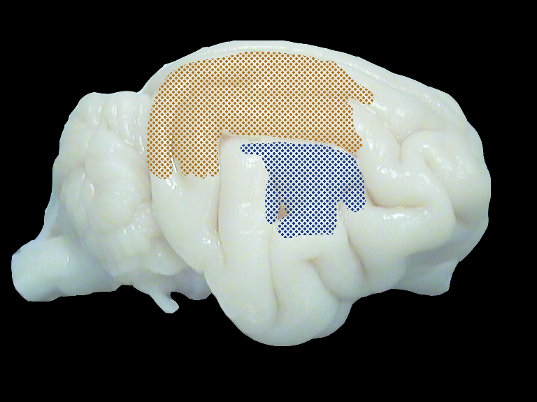
Cat brain. Shown are the direct neighbouring visual and auditory zone (DZ) along the suprasylvian sulcus. Recording in both sides of the sulcus in order to study cross-modal plasticity in the congenitally deaf brain.
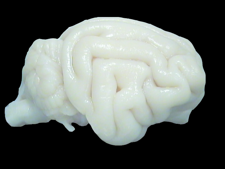
Importantly, we identified a region of interest in the cat cortex, the border zones between visual and auditory cortex within the suprasylvian sulcus. Here the interactions, and especially effects of multisensory integration are likely to provide answer on the differentiation of sensory areas, and the factors influencing the occurrence of cross-modal plasticity in the deaf cortex.
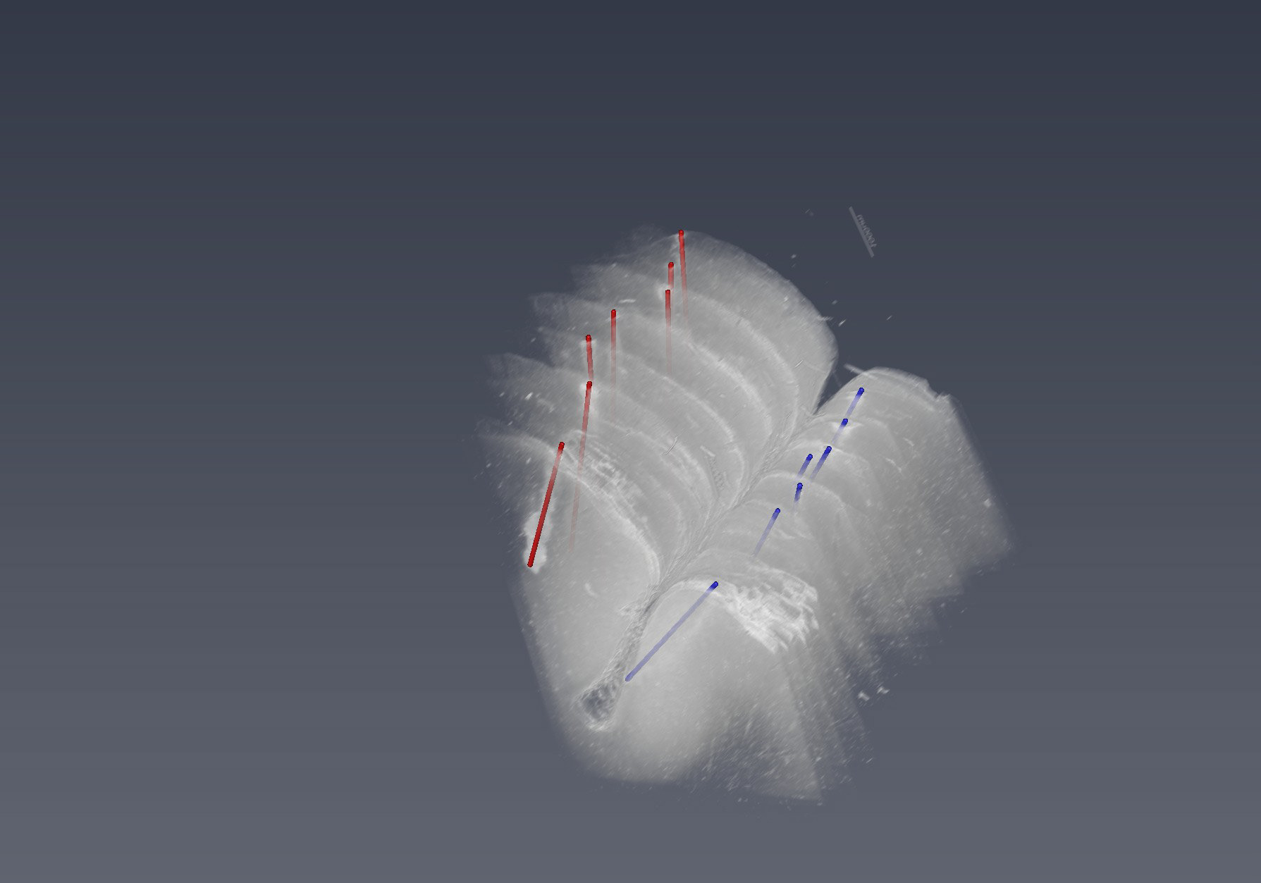
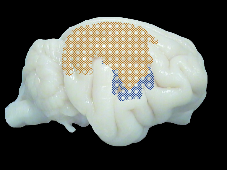
The reconstruction of electrode tracks in the cat brain along the suprysylvian sulcus. Electrode tracks were reconstruced from histological slices followed by a 3D reconstruction.
We have established monaural and bilateral implantation in hearing and congenital deaf cats, and approaches to assess the lower auditory pathway structures by ABR and EBR testing, important for assessment of CI functionality (Land et al. 2016).
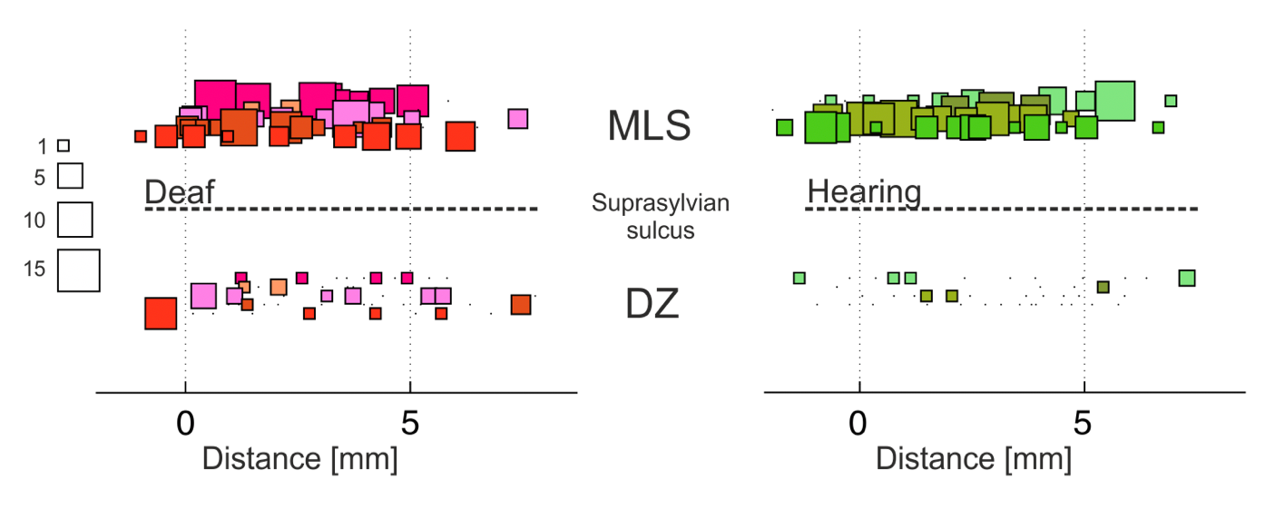
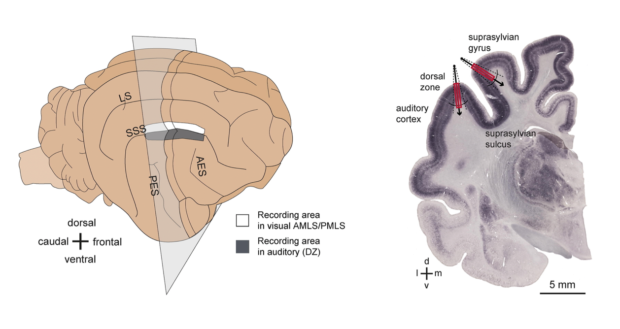
Comparison of visual evoked responses in the congenitally deaf and hearing brain. Squares show the strength of activity on the visual (top) and auditory (bottom) side of the suprasylvian sulcus. In the congenitally deaf brain, visual responsiveness is increased in the auditory cortex, indicating cross-modal plasticity.
References
Land, R. et al. Cross-Modal Plasticity in Higher-Order Auditory Cortex of Congenitally Deaf Cats Does Not Limit Auditory Responsiveness to Cochlear Implants. J. Neurosci. 36, (2016).
Land, R., Radecke, J.-O. & Kral, A. Congenital Deafness Reduces, But Does Not Eliminate Auditory Responsiveness in Cat Extrastriate Visual Cortex. Neuroscience 375, 149–157 (2018).
Kral, A., Yusuf, P. A. & Land, R. Higher-order auditory areas in congenital deafness: Top-down interactions and corticocortical decoupling. Hear. Res. 343, 50–63 (2017).